Revolutionizing Blood Vessel Imaging: The Breakthrough of UI-VISA in Vascular Image Segmentation
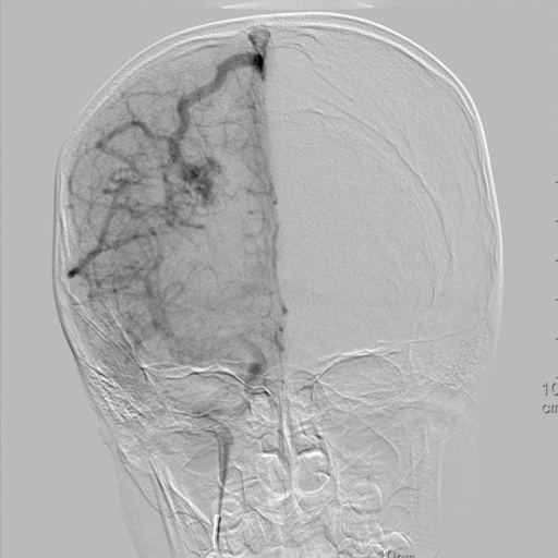
Researchers at the University of California, Merced, have made significant strides in the field of medical imaging with their innovative approach to vascular image segmentation, detailed in their recent paper on UI-VISA (U-Net Initialized Vascular Image Segmentation Architecture). This hybrid architecture uniquely combines deep learning techniques with traditional image processing methods to enhance the accuracy and reliability of imaging vascular structures in digital subtraction angiography (DSA) scans.
Challenges in Traditional Vascular Imaging
Accurate segmentation of vascular structures is critical for diagnosing various cerebrovascular diseases, but traditional methods often face significant challenges. Standard approaches like U-Net can produce fragmented or disjointed predictions, particularly in thinner and branching vessels. Manual tracing, while accurate, is tedious and susceptible to human error. These limitations prompted the research team to develop UI-VISA—a solution that enhances connectivity and continuity in vascular imagery.
The UI-VISA Advantage
UI-VISA presents a notable advancement by leveraging the strengths of both U-Net’s powerful segmentation capabilities and the region growing algorithm’s (RGA) focus on connectivity. Initially, UI-VISA employs a modified U-Net to predict vascular structures. These predictions then serve as informed seed points for a CNN-guided RGA, which refines the segmentations by enforcing local continuity and recovering the fine details often overlooked by conventional deep learning models.
Methodology: A Hybrid Approach
The hybrid nature of UI-VISA is its standout feature. The authors explain that by combining a learned model (U-Net) with a region growing algorithm, UI-VISA effectively enhances segmentation performance. This method not only improves accuracy but also resolves some of the inefficiencies of traditional methods that rely on random seed point initialization, which can lead to computationally expensive errors.
In rigorous testing, UI-VISA achieved the highest scores on critical metrics compared to standalone U-Net and previous region-growing frameworks. For instance, UI-VISA's mean Dice and clDice scores indicated a statistically significant improvement (p = 0.023), showcasing its ability to preserve the intricate connectivity characteristic of blood vessels.
Results and Future Implications
The research findings demonstrate that UI-VISA not only outperforms existing approaches in segmentation accuracy but also significantly cuts down the processing time compared to prior methods. While still slower than the U-Net alone, UI-VISA presents a 3-5 fold faster performance than traditional region growing algorithms. This balance of speed and accuracy is crucial in clinical settings where timely and reliable imaging contributes to better disease management.
As technology continues to advance, the implications of UI-VISA could extend beyond imaging blood vessels, potentially offering a foundation for further innovations in medical diagnostics and treatment planning across various imaging modalities. The integration of deep learning and classic image processing techniques reflects a promising direction for the future of medical imaging, emphasizing efficiency and precision.
With the successful development and validation of UI-VISA, the future of vascular imaging looks brighter, offering clinicians improved tools for diagnosing and managing life-threatening vascular diseases with greater confidence.
Authors: Asees Kaur, Suzanne S. Sindi, Erica M. Rutter