Revolutionizing Pancreatic Cancer Surgery: How Deep Learning Could Change Resectability Assessments Forever
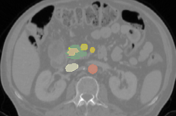
In the high-stakes arena of pancreatic cancer treatment, where timely and accurate surgical decisions can mean the difference between life and death, researchers have unveiled a groundbreaking innovation. A new study proposes a fully automated, multimodal deep learning framework that could transform how surgeons determine whether a patient is eligible for surgery. This innovative approach not only analyzes intricate 3D CT images but also combines them with crucial clinical data to categorize patients into three resectability groups defined by the National Comprehensive Cancer Network (NCCN).
Why Resectability Matters
Pancreatic ductal adenocarcinoma (PDAC) is notorious for its poor survival rates; less than 10% of patients survive five years post-diagnosis. Accurate assessments of a tumor's resectability—meaning whether it can be surgically removed—are vital. Currently, expert evaluations often differ widely, with inter-observer agreement sometimes falling below 70%. This variability can lead to inconsistent treatment decisions, making it imperative to find more reliable methods for evaluating operability.
The Deep Learning Solution
The research team conducted a study on a cohort of 159 patients, utilizing deep learning to fuse information from 3D contrast-enhanced CT scans with 17 structured clinical variables, such as age, BMI, and comorbidities. Their system achieved impressive performance: an accuracy rate of 85% and an Area Under the Curve (AUC) of 0.86 for classifying patients into resectable categories. This remarkable accuracy surpasses many current methods that rely solely on expert analysis or single-source imaging.
Anatomy Meets Clinical Data
Central to this approach is the use of a Swin-UNETR backbone, a novel deep learning model that benefits from both anatomical outlines of the tumor and pertinent clinical factors. By incorporating anatomical supervision during training, the system learns to discern vital structures like major blood vessels and effectively separates tumor areas during analysis. This anatomical awareness enhances diagnosis accuracy without requiring cumbersome segmentation masks during routine clinical use.
Real-World Applications and Future Prospects
The study confirms the implementation's efficacy, not only within the institution where it was developed but also in external validations across different hospitals, thus underscoring its potential for broad clinical application. The researchers emphasize the need for further assessments to test the model's effectiveness in larger, diverse patient groups and to refine its interpretability further.
Conclusion
This pioneering multimodal deep learning framework could significantly improve the decision-making process surrounding pancreatic cancer surgeries. By leveraging advanced imaging and clinical data, the model sets a high standard for future studies aimed at bettering the landscape of oncological practices. As the researchers prepare to share their methods publicly, including data and model weights, the community watches eagerly for the implementation of this cutting-edge technology in clinical settings.
As we stand on the precipice of a new age in medicine, innovations like these might pave the way for faster, more accurate, and ultimately life-saving decisions for patients battling pancreatic cancer.
Authors: Vincent Ochs, Christoph Kuemmerli, Florentin Bieder, Julia Wolleb, Joël L. Lavanchy, Julia Ruppel, Jan Liechti, Stephanie Taha-Mehlitz, Christian Andreas Nebiker, Beat Müller, Giuseppe Kito Fusai, Joerg-Matthias Pollok, Anas Taha, Philippe C. Cattin, Sebastian Staubli