Transforming Infant Brain Imaging: BabySeg’s Groundbreaking Multi-Modal MRI Segmentation
In a remarkable leap for pediatric neuroimaging, researchers from Massachusetts General Hospital and MIT have unveiled BabySeg, a revolutionary deep learning framework designed to accurately segment infant brain scans from diverse MRI protocols. This innovation addresses critical challenges in analyzing brain development during early childhood, a stage characterized by rapid anatomical changes and difficult imaging conditions.
The Challenge of Infant Brain Imaging
Accurate segmentation of MRI scans is essential for understanding human brain development, particularly in infants and young children. Transitioning from general population imaging to specialized infant protocols has presented obstacles due to the smaller size of infant anatomy, varied imaging techniques, and frequent motion artifacts. These factors complicate efforts to capture high-quality images necessary for reliable analysis, leading to a proliferation of narrowly focused segmentation models that often fall short in clinical applications.
Introducing BabySeg
BabySeg is a novel segmentation framework that stands out by integrating a flexible group convolutional network with an innovative training strategy. This system can accommodate a wide variety of MRI contrasts, enabling it to generalize across different imaging captures without the need for retraining the model. By doing this, BabySeg allows for improved and consistent segmentation across different age groups, from extremely preterm infants to those up to five years old.
How BabySeg Works
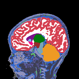
At the core of BabySeg's design is its ability to process multiple input modalities simultaneously—this means it can effectively handle various MRI scans taken during the same session or across different sessions. The model employs domain randomization techniques, which create diverse synthetic training data to enhance its performance in real-world scenarios. As a result, BabySeg can tackle the challenges posed by inconsistent imaging quality and variations in brain development without compromising accuracy.
Remarkable Results
In practical applications, BabySeg demonstrated state-of-the-art performance, achieving segmentation accuracies that not only compare favorably against existing models but do so in significantly less computational time—up to 24 times faster in some cases. This efficiency could translate to faster clinical workflows and improved care for patients. Additionally, the model’s robustness allows it to maintain performance even when exposed to scans with unexpected variations, such as those found in clinical settings.
The Future of Pediatric Neuroimaging
The introduction of BabySeg holds significant implications for researchers and clinicians alike, particularly in studying developmental disorders and brain maturation. By providing a versatile, fast, and more accurate segmentation framework, BabySeg paves the way for future research and advances in pediatric neuroimaging, potentially transforming how we understand and assess brain health in young children.
Research conducted by Malte Hoffmann, Lilla Zöllei, and Adrian V. Dalca, underscores the importance of innovation in medical imaging, and their findings are set to reshape pediatric imaging standards and methodologies.

