Unlocking the Secrets Behind Cell Defects: How Geometry and Obstacles Shape Cellular Behavior
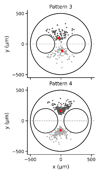
Understanding how cells organize and interact is crucial for advancing fields like tissue engineering and regenerative medicine. A recent research study led by Mina Kamao and colleagues from the University of Tokyo has unveiled a fascinating connection between the geometry of cell environments and the stable configurations of topological defects in cellular populations. This research focuses on how the presence and size of obstacles within cell-cultured domains can significantly influence cell alignment and stability of defect structures.
The Importance of Topological Defects
Topological defects are specific irregularities in the arrangement of cells and molecules, much like imperfections in a crystal lattice. In biological tissues, these defects can play crucial roles, affecting how cells behave and interact. The study highlights the significance of nematic order in confined cell populations, which governs the symmetry and stability of these defects.
Negative Euler Characteristics: A Game-Changer
The research primarily delves into environments characterized by negative Euler characteristics—essentially defining domains with multiple holes or obstacles. Previous studies mainly concentrated on domains with positive characteristics, but this study shifts focus to the unique dynamics present in more complex geometries. Here, scientists discovered that the size and configuration of internal obstacles led the formation of stable pairs of defects characterized by a charge of −1/2.
Experimental Observations Unraveled
The researchers conducted experiments with C2C12 mouse myoblasts, employing microwell substrates that included varying sizes of circular obstacles. Notably, they found that the most stable configurations occurred when these obstacles were sufficiently large, leading to a prevalence of two −1/2 defects within the system. Time-lapse imaging allowed researchers to observe the dynamic nature of cell behavior, revealing insights into how the cellular alignment interacts with surrounding structures.
Predicting Defect Configurations
To validate experimental findings, the researchers devised a theoretical framework based on the physics of nematic liquid crystals. By employing a detailed analysis of Frank elastic energy—a measure related to the stability of defect configurations—scientists successfully predicted how these defects would align and position themselves relative to the obstacles. The theoretical predictions matched experimental results within a remarkable range of accuracy, highlighting the effectiveness of the proposed model.
Implications for Understanding Morphogenesis
This breakthrough carries significant implications for understanding morphogenesis—the process by which cells and tissues develop and organize. The study suggests that cellular tissues may actively control their alignment based on the geometry of their environment, influencing various biomechanical phenomena. As such, this research paves the way for future studies on how manipulating tissue geometries could lead to enhanced tissue engineering strategies and improved regenerative therapies.
In conclusion, this investigation reveals a fundamental relationship between cellular behaviors and their geometrical contexts. By continuing to explore how topological defects and internal structures interact within cellular populations, researchers can unlock new pathways for biological innovation.